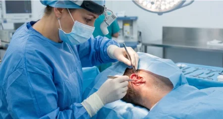

What Is Revision Otoplasty?
Revision Otoplasty is a secondary ear surgery performed to improve structural, cosmetic, or healing-related concerns following a previous otoplasty. The procedure may address recurrent prominence, asymmetry, cartilage irregularities, visible scars, overcorrection, or an unnatural ear position.
Secondary surgery is often more complex than primary otoplasty because the original anatomy has already been altered. Scar tissue, previous cartilage removal, permanent sutures, and changes in tissue flexibility can influence the available correction.
Careful evaluation is therefore essential before another operation. The objective is usually to create better balance and more natural contours rather than attempting to achieve perfect symmetry between the ears.
Why Is Revision Otoplasty Performed?
Patients may consider Revision Otoplasty when the outcome of an earlier operation remains unsatisfactory or changes over time.
One ear may gradually become more prominent, cartilage folds can appear excessively sharp, or the ears may sit too close to the scalp. Other concerns include visible sutures, irregular contours, scar problems, or differences between the two sides.
Revision surgery can also become necessary after complications such as cartilage distortion or wound-healing problems.
The reason for dissatisfaction should be clearly identified before treatment because each problem requires a different corrective strategy.
Common Reasons for Unsatisfactory Otoplasty Results
Unsatisfactory results can develop from recurrence, overcorrection, undercorrection, asymmetry, irregular cartilage folding, excessive conchal reduction, scar formation, or changes in fixation.
Some problems become noticeable during early healing, while others develop gradually as swelling resolves and cartilage responds to its new position.
A previous Revision Otoplasty assessment should distinguish temporary postoperative differences from established structural problems. Mild asymmetry during early recovery does not necessarily indicate a failed procedure.
Long-term concerns should be evaluated after sufficient healing whenever medically appropriate.
Who Is a Good Candidate for Revision Ear Surgery?
Suitable candidates for Revision Otoplasty generally have a persistent concern from previous ear surgery that can reasonably be improved with another procedure.
Candidates may have recurrent prominence, obvious asymmetry, unnatural folds, visible scars, overcorrected ears, or cartilage irregularities. Patients should also be healthy enough for surgery and understand the limitations created by previous operations.
The amount and quality of remaining cartilage are particularly important. Multiple previous surgeries can reduce available tissue and make reconstruction more complicated.
Realistic expectations are essential because secondary surgery may improve existing problems without completely restoring untouched ear anatomy.
When Can Revision Otoplasty Be Performed After the First Surgery?
The timing of Revision Otoplasty depends on the problem being treated and how far healing has progressed.
Swelling, firmness, scar tissue, and cartilage position continue changing for months after primary surgery. Operating too early can make it difficult to distinguish temporary healing changes from permanent structural concerns.
For non-urgent cosmetic revisions, surgeons often prefer to allow several months for tissues to stabilize before making a final decision.
However, complications such as infection, hematoma, exposed cartilage, or significant wound problems may require earlier medical intervention. The appropriate timing must therefore be determined individually.
How Is a Secondary Otoplasty Procedure Planned?
Planning Revision Otoplasty requires detailed analysis of both the current ear shape and the changes created during the original operation.
The surgeon evaluates ear projection, antihelical folds, conchal anatomy, lobule position, cartilage thickness, scars, skin condition, and symmetry. Previous operative information can also help identify cartilage removal and suture placement.
Photographs from before the first procedure may provide useful information about the original anatomy.
A secondary operation should target specific structural problems rather than automatically repeating the original technique. Each ear may require a different correction.
Correcting Ear Asymmetry with Revision Otoplasty
Revision Otoplasty can improve ear asymmetry when differences result from unequal cartilage folding, projection, fixation, scar contraction, or recurrence after previous surgery.
Correction may involve releasing an overcorrected area, strengthening a weak fold, changing suture placement, or reshaping cartilage. One ear may require considerably more treatment than the other.
Natural differences in skull shape, ear size, and cartilage structure can limit perfect symmetry.
The realistic objective is improved visual balance from several angles while maintaining natural ear contours and avoiding excessive correction.
Treating Recurrent Prominent Ears After Otoplasty
Recurrent prominence can occur when cartilage gradually moves toward its previous position or when surgical fixation does not remain sufficiently stable.
During Revision Otoplasty, the underlying reason for recurrence should be identified before correction. Strong cartilage memory, weakened sutures, inadequate initial reshaping, or healing characteristics can contribute to the problem.
Treatment may involve renewed cartilage reshaping, additional fixation, or modification of the conchal region.
The correction should remain controlled. Pulling a recurrent ear excessively close to the scalp can replace one aesthetic concern with an unnatural pinned-back appearance.
Cartilage Correction in Revision Ear Surgery
Cartilage management is one of the most important aspects of Revision Otoplasty because previous surgery may have already changed cartilage thickness, strength, and shape.
The surgeon may need to reshape existing cartilage, release unnatural folds, reposition cartilage segments, or reinforce areas that lack adequate support.
In more complicated cases, previous cartilage removal can make reconstruction challenging. Additional tissue support may occasionally be considered depending on the defect and available anatomy.
Conservative correction is particularly important during secondary procedures because repeatedly weakening cartilage can increase the risk of irregular contours.
Scar Management During Revision Otoplasty
Scar tissue can influence both the appearance and technical difficulty of Revision Otoplasty.
Previous incisions are often located behind the ear, but scars may become widened, raised, thickened, or irregular. Internal scar tissue can also restrict cartilage movement and make surgical dissection more difficult.
When appropriate, an existing scar may be revised during secondary surgery. However, removing a scar creates a new healing incision and cannot guarantee an invisible final scar.
Patients with previous hypertrophic or keloid scarring require particularly careful evaluation because they may remain at increased risk of abnormal scar formation.
Recovery After Revision Otoplasty
Recovery following Revision Otoplasty usually includes swelling, bruising, tenderness, tightness, and temporary changes in sensation around the ears.
A protective dressing or headband may be recommended during early healing. Patients are commonly advised to avoid direct pressure on the ears and may need to sleep on their back temporarily.
Visible swelling often improves significantly during the first few weeks, although secondary procedures can sometimes produce more prolonged firmness because scar tissue has already formed from previous surgery.
Exercise, contact sports, and activities that could bend or strike the ears should only be resumed according to postoperative instructions.
What to Expect During the Healing Process
Healing after Revision Otoplasty occurs gradually, and early ear position should not be interpreted as the final result.
During the first days, swelling can make the ears look uneven, overly close to the head, or different in size. Bruising and tenderness may also temporarily affect appearance.
As swelling decreases, the new cartilage contours become easier to evaluate. Internal scar tissue continues softening and reorganizing for several months.
One ear can heal faster than the other. Minor temporary differences are therefore common and do not automatically indicate persistent postoperative asymmetry.
Revision Otoplasty Results and Ear Appearance
The goal of Revision Otoplasty is to improve existing ear concerns while producing smoother contours and a more balanced relationship between the ears and head.
Results depend heavily on the condition of the cartilage and skin after previous surgery. Patients with limited scarring and adequate remaining cartilage may have different corrective possibilities from those who have undergone multiple operations.
Complete symmetry cannot be guaranteed because ears naturally differ and previously operated tissues may respond differently during healing.
A successful result should look balanced without eliminating the natural folds, curves, and projection that give the ear its normal three-dimensional appearance.
How Long Do Revision Otoplasty Results Last?
Results from Revision Otoplasty are intended to provide long-term structural improvement once the cartilage and surrounding tissues have healed.
Repositioned cartilage can remain stable for many years. However, secondary surgery cannot guarantee that ear position will remain completely unchanged throughout life.
Cartilage elasticity, scar contraction, trauma, fixation stability, tissue aging, and individual healing characteristics can influence long-term appearance.
Recurrence remains possible, particularly when strong cartilage memory contributed to failure of the first operation. Careful correction of the underlying structural cause can help improve long-term stability.
Revision Otoplasty Prices 2026
Revision Otoplasty Prices 2026 vary considerably because secondary ear surgery must address the individual changes created by previous procedures.
Is revision otoplasty more difficult than primary otoplasty?
Revision otoplasty can be more technically challenging than primary ear surgery because the cartilage and surrounding tissues have already been altered. Scar tissue, previous sutures, cartilage removal, and changes in tissue flexibility can make normal surgical planes more difficult to identify.
How long should I wait before having revision otoplasty?
The appropriate waiting period before revision otoplasty depends on healing progress and the reason another procedure is being considered. Swelling, scar firmness, cartilage position, and asymmetry can continue changing for several months after the first operation.
Can prominent ears return after otoplasty?
Prominent ears can partially return after surgery if the corrected cartilage gradually moves toward its previous position. This recurrence can happen because ear cartilage naturally has elasticity and structural memory.
Can revision surgery improve uneven ears?
Persistent differences between the ears can often be improved with revision otoplasty when asymmetry results from cartilage position, unequal projection, irregular folds, scar contraction, or different responses to the original surgery.
Does revision otoplasty leave additional scars?
Revision otoplasty can create additional scar tissue because another surgical procedure requires reopening or creating incisions. Whenever possible, existing scars behind the ear may be used to access the cartilage and reduce the need for additional visible incisions.
How long does swelling last after revision ear surgery?
Swelling after revision otoplasty is usually most noticeable during the first several days and gradually decreases during the following weeks. The exact duration varies according to the amount of cartilage reshaping, scar-tissue dissection, and reconstructive work performed.